

Definition
A fracture of the proximal femur also named hip fracture, consists in the break of the upper segment of the femur bone in proximity of the pelvic socket, the acetabulum.

Pathology
A proximal femur fracture is a serious condition that requires immediate management in the Emergency Department. It is a serious injury because it frequently occurs in elderly people and may lead to life threatening complications, like pneumonia or deep venous thrombosis. Nowadays, the incidence of proximal femur fractures is increasing as a consequence of prolonged life expectancy. In the elderly the loss of bone calcium or osteoporosis, which reduces bone density, combined with a higher risk of falls are a major medical concern. In the younger population a proximal femur fracture requires a high energy traumatic impact.

Classification
Proximal femur fractures can occur in different locations and are generally divided into:
Intra-capsular (fracture of the femur within the joint capsule)
Extra-capsular (fracture of the femur outside the joint capsule).
There are three types of proximal femur fractures:
Femoral neck fracture or trans-cervical fracture, proximal to the trochanters
Trochanteric femur fracture or inter-trochanteric fracture, between the greater trochanter and the lesser trochanter
Femoral neck or sub-trochanteric fracture located distally, beneath the trochanters
The fracture to the proximal femur is displaced when the bone extremities have lost their anatomical alignment or non-displaced when the break does not separate the femur bone segments. A displaced fracture requires a reduction into the correct position whereas in a non-displaced fracture, reduction is not necessary.

Causes
In older individuals a fall is the most frequent cause for a proximal femur fracture (approximately of 90% of all hip fractures). Even a fall from a standing position can lead to a proximal femur fracture in this patient group. A fall can be the consequence of medical conditions including limited vision, balance problems, sudden loss in blood pressure and heart arrhythmia causing people to faint.
A stress fracture of the proximal femur in the elderly is due to bone weakening by osteoporosis. This type of fractures does not require a traumatic impact or a fall to occur. Women with an average age of 80 years are particularly vulnerable to a proximal femur fracture having an 8 to 10 ratio of incidence compared to men. In the younger population a proximal femur fracture is rare and arises from high energy trauma such as car accidents or falls from a significant height.

Risk factors
Leading risk factors for a proximal femur fracture include:
Elderly age
Female gender: women lose bone density after menopause
Osteoporosis
Chronic medical conditions (hyper-/hypotension, stroke, heart arrhythmia, thyroid dysfunction)
Medications e.g. steroids weaken bone density or relaxants facilitating falls
Lack of physical fitness
Use of tobacco and alcohol
Car accidents
Working at height (carpenters, electricians, builders, painters)
Symptoms
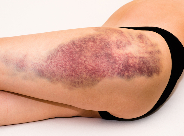
A fractured proximal femur causes an immediate, severe pain. The patient is likely unable to move, stand up and walk. As a result of a proximal femur fracture the leg of the side affected is positioned at a different angle directed outwards and may appear shorter. On the hip side of the fall the patient may present a bruise, swelling and stiffness.

Diagnosis
Given the serious nature of a proximal femur fracture the first diagnosis is normally executed at the Emergency Department. Following admittance to the hospital, the patient is firstly treated with pain killers (analgesics) and fluid replacement, prior to begin radiologic evaluation with X-rays. Often a CT scan or an MRI is performed to characterise the fracture type and the potential involvement of the cartilage and ligaments of the hip joint. After image assessment, the orthopaedic surgeon will opt for the best treatment appropriate to the type of fracture and the general condition of the patient.
Treatment

Nonoperative treatment
A non-displaced, intracapsular proximal femur fracture is normally treated conservatively but requires a longer hospitalisation period. Nonoperative treatment of a proximal femur fracture increases the risk of delayed bone displacement and involves frequent monitoring by X-ray. In complex proximal femoral fractures, surgery is avoided only if the patient has medical conditions that pose a high risk of complications.

Surgical treatment
The guidelines from the National Institute for Health and Clinical Excellence (NICE) recommend that surgery for a proximal femur fracture is carried out within one or two days from the accident. Prior to surgery the patient is treated with analgesic drugs for pain relief.
Non-displaced fractures - Surgery via internal fixation may necessary for the treatment of non-displaced intra-capsular proximal femur fracture to consolidate the bone fragments. A number of metal devices are available for this surgery such as screws, nails, plates and rods. This procedure ensures a superior healing by maintaining a proper bone alignment with the advantage to accelerate the general recovery of the patient allowing early mobilisation.
Displaced fractures - A displaced intra-capsular proximal femur fracture, firstly involves an open reduction to re-align the bone fragments followed by internal fixation with the application of a plate alongside the femur bone fixed with screws. Intra-medullary nailing involves the insertion of a long nail through the femoral canal containing the bone marrow. The nail is fixed at the upper part of the rod through the femoral neck into the femoral head.
A hip replacement or arthroplasty is necessary if the fracture has severely compromised the integrity of the bones. With this method the damaged bones are firstly removed and then replaced with artificial prostheses. This includes a total hip arthroplasty, with replacement of both the femur head and the acetabulum especially if the patient suffers from a displaced intra-capsular hip fracture and chronic osteoarthritis. Hemiarthroplasty involves only the replacement of the femoral head and part of the shaft.
Extra-capsular proximal femur fracture - Surgery utilises various devices such as a sliding hip screw or an intra-medullary nail as mentioned earlier for displaced intra-capsular fractures.

Complications
Complications that may occur following a proximal femur fracture include:
Local infection - requiring antibiotic treatment as a prevention measure
Deep vein thrombosis (DVT) and pulmonary embolism - thrombosis mostly occurs in the deep veins of the lower limbs due to prolonged immobility. Patients are usually treated with prophylactic administration of anti-clotting medications (warfarin, aspirin)
Blood loss - this can occur during or after surgical treatment. Rarely, a blood transfusion is needed
Fracture nonunion - occurs when the fracture does not heal completely or when the healed bone presents altered anatomical alignment.
Avascular necrosis - in case of an intra-capsular hip fracture the bone break may injure the vessels supplying the femoral head causing the death of the bone tissue. Avascular necrosis of the femoral head can lead to chronic pain of the hip region.
Pressure ulcers - due to extended immobilisation and the fragile skin in the elderly, the pressure of the weight on specific areas of the body can form skin ulcers. This can be prevented using specific bed characteristics, massage and frequent reposition of the patient.When pressure ulcers become infected they need immediate antibiotic treatment
Pneumonia - used to be the main cause of death in older patient that were immobilised for several weeks. Nowadays surgical treatment is more often delivered to aged patients accelerating their mobility and reducing the risk of infections
Urinary tract infection - is another complication due to immobilisation, hygiene and insufficient hydration

Rehabilitation
Particularly in elderly patients it is critical to begin rehabilitation immediately after surgery through gradual and assisted walking to prevent medical complications. This phase may require the patient to be admitted to a cared facility or receive regular in-home visits by an occupational therapist to return to independent daily living activities.
A wheelchair, crutches, walking stick, or a walker may be necessary to support the patient during the first weeks up to 12 months after a proximal femur fracture while recovering in function. Physiotherapy during the first 6-12 weeks post surgery include:
Ice/heat treatment
Antiinflammatory therapy
Exercise to strengthen quadriceps, hamstrings and gluteal muscles
Hydrotherapy
Massage
Joint mobilisation
Guided return to activity
Use of high chairs and walking devices
Weight loss in overweight patients

Prevention
A proximal femur fracture poses a higher risk for a second such a fracture. Therefore prevention is key to avoid recidivism. In osteoporotic patients treatment includes the administration of the bisphosphonate group of drugs with additional supplements of calcium and vitamin D.
Other preventive measures include:
Exercise and maintenance of muscular strength in the elderly (walking, swimming)
Use of supporting walking devices in the elderly
Wearing of hip protectors
Adherence to occupational health and safety procedures and road traffic safety equipment (seatbelt, harness, balustrades)
Monitoring of chronic diseases and pharmacological use (blood pressure medications)
Alcohol and drug rehabilitation
Quit smoking (impairing bone healing)
Removal of carpets or other items facilitating falls
Modify habits (use laced shoes, illuminate house at night, install railings, non-skid tiles in bathroom).