

Definition
Fractures of the pelvis consist in the break of any bones forming the pelvic ring possibly involving portion of the lower spine, the sacrum and tailbone (coccyx).
Pathology
The fracture of the pelvis is frequently the consequence of high energy force that impacts directly on the pelvic ring or occur via a muscle strain resulting into avulsion fractures. Occasionally stress fractures arise in older individuals due to poor bone quality or following inappropriate training. Pelvic fractures represent 3% of all fractures with an incidence of 1-2% of fractures in children.
The most common types of pelvic fractures include single pubic rami and avulsion fractures. Fractures of the pelvis may involve the pubis, ilium and ischium. The severity of a fracture is determined by the concomitant injury to the digestive and reproductive organs enclosed in the pelvic cavity. Pelvic fractures can lead to damage of large arteries and veins causing severe blood loss as well as nerve injuries. For this reason they require immediate medical care at the Emergency Department.

Classification

Various classification systems are available to define fractures of the pelvis, whereby the Tile is more broadly used than the Apley or the Young classifications.
Tile classification
relies on the status of the posterior sacroiliac complex:
Type A:
A1 Stable injuries including: avulsion fractures, isolated pubic rami fractures, iliac wing fractures
A2 single stable fractures of the pelvic ring. Avulsion fractures occur in the region where muscles attach to pelvic bones:
Anterior inferior iliac spine: rectus femoris muscle
Anterior superior iliac spine: Sartorius muscle
Ischial tuberosity: hamstrings
Type B: These fractures are rotationally unstable but vertically stable.
B1: also named 'open book’ is a fracture caused by antero-posterior compression, which separates the symphysis and broadens the sacroiliac joints
B2: ipsilateral compression results in fractures of the pubic bones and their overlap
B3: contralateral compression injury causes a fracture of the pubic rami on one side and a compression sacroiliac injury to the opposite side
Type C: These most severe pelvic fractures are rotationally and vertically unstable and often associated with extensive blood loss and high mortality. The pelvic ring is completely disrupted or dislodged at two or more regions. They are further divided into:
C1: unilateral
C2: bilateral
C3: concomitant to acetabular fracture.

Young classification
is based on the mechanism of injury: lateral compression, antero-posterior compression, vertical shear or a combination of these forces.
Lateral compression (LC) fractures produce transverse fractures of the pubic rami, located either to the same side (ipsilateral) or to the opposite side (contralateral) of an injury to the posterior portion of the pelvis. According to the Young classification, fractures of the pelvis are divided into three grades depending on their complexity:
Grade I - Associated with sacral compression on side of impact
Grade II - Associated with posterior iliac ("crescent") fracture on side of impact
Grade III - Associated with contralateral sacro-iliac joint injury

Apley classification
divides pelvic fractures into:
Avulsions caused by high force action of the following muscles:
Sartorius at the anterior-superior iliac spine (ASIS), the anterior extremity of the iliac crest
Rectus femoris at the anterior-inferior iliac spine (AIIS), the upper lateral parts of the pelvis
Adductor longus at the pubis
Hamstrings at the ischial tuberosity

Causes
The major causes leading to pelvis fractures in adults are:
Falls/falls from height more frequent in elderly (8-10%)
Crush injuries (3-6%)
High-velocity impacts in motor vehicle accidents
Osteoporosis
Inappropriate training
Muscle and joint weakness
In children:Pedestrians hit by motor vehicle (60-80%)Motor vehicle accidents (20-30%)

Risk factors
The main factors for a pelvic fracture are:
Driving motor vehicles and motorbikes
Working at height
Osteoporosis and balance problems in the elderly
Sport (muscle strains leading to avulsion fractures)
Thrill seeking activities (planking)
Mining
Blast injury and penetrating trauma in the military

Symptoms
In addition to the typical symptoms consequent to a pelvic fracture such as pain, swelling and bruising the patient may present manifestations indicative of injuries to the abdominal organs, vessels and nerves:
Tenderness at touch
Pelvic instability
Inability to move or reduced range of movement
Haematuria (blood in urine)
Rectal and/or vaginal bleeding
Neurological deficit of lower extremities
In more severe cases, damage to internal organs may lead to persistent shock or loss of vital signs:
Low blood pressure < 90 mm HG
Pulse >100
Central venous pressure (CVP) < 5 cm with a water manometer
Urine output < 30ml/hour (despite fluid replacement)
Absence of vital signs torrential haemorrhage >12 units of blood/2 hours
%20%20copy.jpeg)
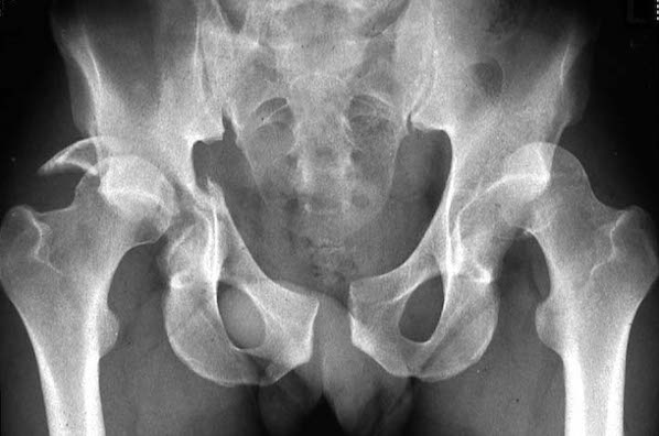
Diagnosis
A pelvic fracture is a serious condition that requires admission to the Emergency Department. The mechanisms of injury may give a good indication for a pelvic fracture. Physical examination includes palpation, monitoring of anatomical changes and a number of instability, functional and neurological tests to determine the presence of abnormalities in the range of movement, potentially underlying a fracture. To exclude the existence of associated injuries, the examiner will also investigate the upper abdominal region as well as the eventuality of a fracture or dislocation of the hip and nerve damage.
In pregnant women the well being of the foetus is mandatory. In case of haemorrhagic shock immediate emergency care with fluid replacement, blood transfusion and cardiovascular stabilisation may be necessary.
In more severe cases medical examination may also include the assessment of:
Blood pressure
HaematocritUrine analysis (haematuria)
Vaginal bleeding in females
Scrotal haematoma in males (Destot's sign)
Digital rectal and vaginal examination
Spine and femur fractures
The final diagnosis is confirmed via a number of imaging techniques targeting both the bony and the soft tissues of the pelvic region:
X-rays basic - used for initial screening - antero-posterior, inlet view, outlet view
Ultrasound - bleeding, internal injuries
CT scans - detects fracture features and bleedings
MRI - more reliable for subtle fractures and fracture classification
Urethrography (injury to urogenital tract)
Arteriography (bleeding, vascular damage)
Cystography (bladder damage)
Treatment

Nonoperative treatment
Immediate emergency care aims at stabilising the patient as soon as possible after hospital admission due to a suspected pelvis fracture. If the fracture is stable and does not present additional complications conservative treatment is sufficient. It involves an initial short period of bed rest followed by the use of crutches for 3-6 months or until the fracture has healed to avoid full weight bearing. Additionally, the patient receives anticoagulation drugs as prophylaxis against deep vein thrombosis (DVT). Further medications include administration of:
Analgesics
NSAIDs at later stage to avoid early bleeding complications

Surgical treatment
When the pelvis fracture is complicated, or displaced and involves life threatening injuries to pelvic organs, surgical treatment is necessary. This approach may follow two stages: an initial stabilisation of the fractured pelvis while implementing resuscitation procedures to restore hemodynamic instability and repair of injured pelvic organs.
This includes a provisional stabilisation of the pelvis with external devices (pelvic clamp or external fixator). This is followed by a permanent surgical fixation 2-3 days later. The initial management includes also:
Fluid replacement
Reverse shock status
Haemodynamic stabilisation
Embolisation of injured vessels
Secondary surgery
Different methods are available for internal fixation of the fractured pelvis relative to the individual characteristics of the fracture and the accompanying internal injuries. Surgery can be performed from the anterior (incision extending from the anterior superior iliac spine to the iliac tubercle) or the posterior side of the pelvis (incision 2 cm lateral to the posterior superior iliac spine). The surgeon will expose the bone by gently pushing away muscles, vessels and nerves.

Open reduction internal fixation (ORIF) is indicated in the following types of pelvis fractures:
Diastasis (widening) of the pubic symphysis > 2.5 cm
Dislocations of the sacro-iliac joint
Displaced fractures of the sacrum
Posterior or vertical displacement of the hemipelvis (>1 cm)
Rotationally unstable injuries of the pelvic ring
Sacral fractures concomitant to unstable pelvic ring
Displaced sacral fractures accompanied by neurologic injury
These are the most common devices used in ORIF:
Anterior plates on pubic bones
Reduction of hemipelvis with pins inserted in the iliac crestIlio-sacral screws
Two-hole plates placed at 90° to each otherIn crescent fractures (sacro-iliac fracture): cortical lag screws (3.5 mm) placed between the pelvic tables from posterior to anterior plus a 3.5-mm plate along the outer table.The fracture healing progress is monitored with X-rays taken at 2, 6, and 12 weeks after surgery.
Complications
Complications of pelvic fracture include:
Deep venous thrombosis due to prolonged immobilisation
Bleeding from fracture or pelvic vasculature
Intrapelvic compartment syndrome
Damage to bladder, urethra, prostate or vagina (higher incidence of urethral injuries in Straddle fractures associated with sacroiliac diastasis)
Sexual dysfunction
Infections from disruption of bowel or urinary system
Chronic pelvic pain, especially with sacroiliac joint injuries
The prognosis after a pelvic fracture presents an overall mortality of 5-20% rising up to 42% in open fractures. The incidence of mortality and morbidity increases with age and is approximately 50% in people > 70 years. In pregnancy there is a risk of foetal loss of 33% or need of caesarean section in up to 40% of women.

Rehabilitation
Following a pelvic fracture an initial phase of bed rest is required. Subsequently, the patient is allowed to use devices such as crutches and walking stick to reduce weight bearing. This phase can last for a few weeks to months depending on the severity of the fracture, whether the patient was subjected to conservative or surgical intervention as well as general health conditions and age. Finally, a physiotherapist will guide the patient towards a gradual increase of weight bearing and exercise. This can continue for a few months. Additional physiotherapy includes:
Massage
Trigger point release techniques
Dry needling
Joint mobilisation
Stretches
Electrotherapy
Strengthening exercises and core stability (Pilates, swimming, cycling, water running)
Guided return to physical activity, work and sport

Prevention
Pelvic fractures can be prevented by avoiding falls from height or falls in elderly people as well as reducing severity of road and work related accidents. This is achieved by adhering to available standard safety measures. They include:
Automobile seat belts, airbags
Safety equipment to reduce risk of falls from high levels at workplace (belt, harness, balustrade)
Administration of hormones, calcium, vitamin D or other treatments for osteoporosis
Improve physical fitness and strength
Walking devices in elderly individuals